Cytomedins. Immunomodulation. 25 years of research
December 29, 2021
Cytomedins are a special group of biologically active substances produced from animal tissues. They were first obtained in the 1970s by Soviet scientists V.Kh. Khavinson and V.G. Morozov. Cytomedins are peptide bioregulators capable of influencing the basic physiological processes of the human body – differentiation and proliferation of cells, and exchange and reproduction of genetic information.
Benefits of Сytomedins include:
- Targeted action,
- High efficiency,
- Organotropy,
- Mild physiological effect,
- Normalization of the metabolism of cell nuclear structures,
- Compatibility with most medications, and methods of treatment,
- Minimum metabolic stress (optimal pharmacokinetics),
- Free from side effects and contraindications.
The History of Cytomedins from the Russian Empire till Our Days
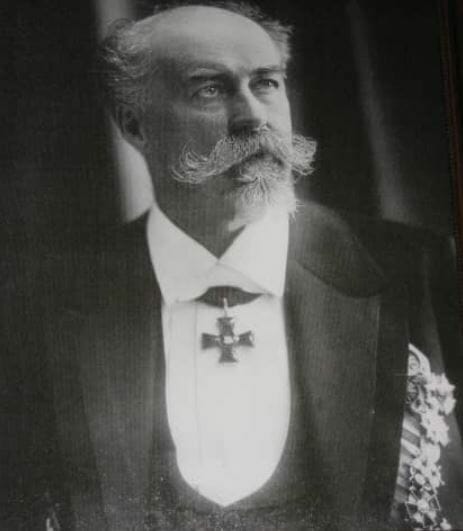
In oriental medicine, the history of the use of animal origin products like soups from bones and powders, etc. is thousand-years old. In European medicine it’s been several centuries. And it probably began at the end of the 18th century when Ch.E. Brown-Séquard (1889, France) made an attempt to use physiologically active substances from the seminal glands as a rejuvenating agent. Next was professor A.V. Poehl (1850-1908, the Russian Empire) who suggested scientific and experimental evidence for the use of animal tissue extracts. He studied the subcutaneous injections of an emulsion from the seminal glands of animals and started the production of organic preparations – spermin and opovarin (see the picture below).


Professor V.S. Gulevich (1900, Russia) isolated “carnosine”, the first of the neuropeptides, a regulator of behavioral reactions that increase the adaptation of cells and tissues to unfavorable conditions. Works of Nobel Prize winners became the background for the use of these drugs: I.I. Mechnikov and P. Ehrlich – theory of phagocytic immunity, Nobel Prize in Physiology and Medicine 1908; D. Watson and F. Crick, M. Wilkinson – discovery of the molecular structure of nucleic acids and its importance in the transmission of information in living matter, Nobel Prize in Physiology and Medicine 1962; F. Jacob and J. Monod – a model of genetic regulation of protein synthesis, Nobel Prize in Physiology and Medicine 1965; P. Berg, W. Gilbert and F. Sanger – fundamental research of the biochemistry of nucleic acids and the determination of the sequence of bases in RNA and DNA, Nobel Prize in Chemistry 1980; A. Ciechanover, A. Hershko and I. Rose – showed the role of short peptides in the transmission of biological information, Nobel Prize in Chemistry 2004. [19]
In the 1970s young researchers from the Leningrad Military Medical Academy (Russia) V.Kh. Khavinson and V.G. Morozov got interested in the physiological properties of the active substances of the peptide nature. They developed a method for obtaining special substances from animal tissues, and called them “Сytomedins”. Later I.P. Ashmarin the academician of the Russian Academy of Medical Sciences proved the fundamental property of regulatory peptides – polyfunctionality, when one peptide can provide regulation of various physiological functions. He also substantiated the cascade principle of action of regulatory peptides. A huge role in the widespread introduction of biologically active peptides into medical practice in Russia belongs to the Saint-Petersburg Institute of Gerontology.
If you have a headache, you need to take analgin. But if you take a course of Cerebramin, a bioregulator from the cerebral cortex, your head will hurt less frequently and less intensely. Bioregulators do not immediately relieve symptoms, but they ensure optimal organ function and thus have preventive function.
V. Kh. Khavinson
Second half of the XX century in biology and medicine is characterized by the rapid development of the studies of the role of peptide bioregulators in the activity of the body. A large number of publications dedicated to the effect of peptides on the course of various physiological functions appear annually.
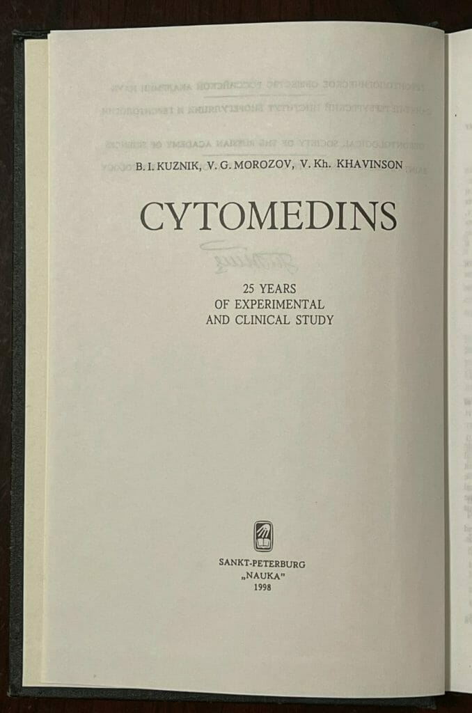
This time we’re sharing with you the information from the monograph called “CYTOMEDINS. 25 Years Of Experimental & Clinical Study” by B.I. Kuznik, V.G. Morozov and V.Kh. Khavinson. The monograph summarizes years of research of the new class of peptide bioregulators that perform the function of humoral carriers of information that is exchanged by cells. In the monograph you will find info about the chemical composition and specific functions of Cytomedins. The authors answer questions about the mechanism of action and their use in practical medicine.
This post can be useful for biohackers, pharmacologists and nootropic enthusiasts who use peptides and other preparations based on Cytomedins. The book has more than 300 pages. But we’ve translated most interesting parts (in our view):
- Introduction
- Composition and Chemical characteristics of Cytomedins
- Specific and Non-specific action of Cytomedins
Introduction [17]
By the beginning of the last decade of the XX century more than 1,000 peptides with an established number of amino acid residues and their sequence of arrangement were isolated from various biological tissues. At the same time, the structure of many peptides has not yet been fully elucidated, although the effects that they stipulate are well studied.
O. A. Gomazkov [20], having analyzed the extensive literature, identifies the following stages in the study of physiologically active peptides:
- The search for new peptides, the study of their physiological effects, tissue localization and chemical structure;
- Identification of new functions of already known peptides;
- Study of factors regulating the physiological activity of certain groups of peptides, ferments of synthesis and degradation, connection with other physiologically active substances and selectivity of receptors;
- Research in the field of molecular “peptidology”;
- Study of the pharmacological regulation of the functions of polypeptides and the synthesis of receptor antagonists and agonists, the search for highly specific enzyme inhibitors;
- Development of computer technologies in the study of peptides, chemical design of peptide fragments, analysis of the molecular topography of active domains of the structure, identification of peptide structures “recognizing” a receptor and “binding” to it, creation of new compounds (peptide analogs, receptor antagonists, peptidase inhibitors) based on these principles.
Polypeptides consisting of separate amino acids and having a relatively low molecular weight (MW) drew close attention after the discovery of neuropeptides – compounds capable of controlling the activity of all organs and systems of the multicellular organism through the central nervous system (CNS). Later it was shown that peptides are found in almost all organs and tissues. It has been established that these compounds, synthesized in the body of higher and lower animals, perform not only a signaling role, but are also directly involved in the regulation of physiological processes, from individual functions of specialized cells to complex behavioral acts. In this regard, many researchers argue that the term “neuropeptides”, proposed in 1969 by De Wied, is outdated and does not reflect the essence and mechanism of action of these compounds.
Biologically active peptide regulators by their nature can be classified as autocoids and cybernins. The former interfere in the course of physiological and biochemical processes directly in the zone where they are formed, the latter exert their influence near the place of synthesis. At the same time, it is known that the same peptide is capable of simultaneously regulating the course of physiological functions both at the site of its formation and far beyond it. Moreover, the effect of the polypeptide can be very diverse.
To date, peptide regulators have been found in the gastrointestinal tract, cardiovascular system, respiratory and excretory organs. With the discovery of the APUD-system (Amine Precursor Uptake and Decarboxylation) it became clear that the gastrointestinal tract contains cells that have secretory functions and produce various biologically active substances of the peptide nature. The very name ‘APUD-system’ suggests that the morphological structures of this group of cells absorb amine precursors and, by carboxylation, convert them into peptide hormones with a broad spectrum of action. In addition, the organs of the digestive system contain virtually all known polypeptides that have ever been found outside of it. These compounds play a significant role in the regulation of the physiological functions of the body, as well as in the development of a number of pathological conditions.
However, later it turned out that similar cells are located in various organs and tissues (respiratory and excretory systems, endocrine glands) that are not related to the digestive apparatus. Therefore the understanding of the APUD-system has significantly extended. It has now been shown that it includes about 40 types of cells. That is why, instead of the term “APUD-system”, another one began to be used more often – the disseminated neuroendocrine system, also called the third nervous system. This name was given to it, because the APUD-system operates autonomously and controls the activity of all internal organs without exception.
Endogenous peptide bioregulators contained in blood, lymph, interstitial fluid and various tissues can have at least three sources of their origin:
1) endocrine tissue cells,
2) neuronal elements of an organ,
3) a depot for the axonal peptide transportation from the CNS.
The last statement is based on the fact that the brain constantly synthesizes, and therefore contains, with a few exceptions, all peptide bioregulators. These facts gave the reason to call the brain a large endocrine organ [7]. It has been established that many peptide bioregulators are capable of entering the periphery via axonal transportation. [13]
In the mid-1980s it was proven that there are information molecules in the cells of human and animal organisms that provide interconnections in the activity of the nervous and immune systems. They were called ‘titins‘. [11] They are formed in the immediate proximity to the corresponding receptive structures. And the distance between the spots of their appearance and application is often comparable to the size of the cell. It has been suggested that titins emerged at the early stages of the formation of biochemical systems of prokaryotes on the basis of ribosomal protein synthesis, exo- and endocytosis processes, and reactions of limited proteolysis. There is no doubt that cellular receptors, like peptide-protein ligands and, first of all, immunoglobulins, can act as pre-hormones, limited proteolysis of which can lead to the appearance of low molecular weight peptide compounds acting as autocrine or paracrine bioregulators. [11]
However, the idea of Cytomedins as a new class of peptide bioregulators was formulated even before the discovery of titins. [6, 4] The term ‘Cytomedins’ comes from the Greek kitos (cell) and the Latin mediator. These compounds provide communication between small groups of cells and thus have a pronounced effect on their specific activity. Apparently, Cytomedins carry certain information from cell to cell, recorded using a sequence of amino acids and conformational modifications. The greatest effect of Cytomedins is manifested in the tissues of the organ from which they are isolated.
Initially, Cytomedins were found in the tissues of the brain, pineal gland and hypothalamus. [2] In addition to the effects on the CNS, these compounds influenced the protective functions of the body and the reproductive organs. Soon, Cytomedins were isolated from the vascular wall. Under their influence, metabolic processes in blood vessels were normalized, and the development of atherosclerosis was prevented. Subsequently Cytomedins were obtained from the primary (thymus, bursa of Fabricius, bone marrow) and secondary (lymph nodes, lymphoepithelial formations, spleen) organs of the immune system. Cytomedins were also extracted from almost all endocrine glands, placenta and its membranes, heart and vascular endothelium, various parts of the gastrointestinal tract, kidneys and liver, bronchi, periodontal tissues, erythrocytes, leukocytes, platelets, blood plasma and serum, lymph, retina, lens and from other organs and tissues.
An important role in the body belongs to peptide growth factors (growth factor of the epidermis, blood vessels, platelets, etc.). They are formed in a wide variety of tissues and not only perform their main function, but also largely determine the activity of various organs and systems.
With the help of the nervous tissue culture, the presence of neurotrophic factors was identified. They are low molecular weight peptides that have a high specificity in relation to neurons of the CNS and the peripheral nervous system. Subsequently, neurotrophic factors were divided into three groups: [16, 8]
- Neurotrophic – supporting the survival and development of neurons,
- Neuritis-stimulating – promoting the growth of neuronal processes,
- Inhibiting neuronal growth.
The most studied neurotrophic factor – nerve growth factor (NGF) – can be obtained in sufficient quantities from the submandibular salivary glands, heart, liver, uterus, placenta and other tissues. At the same time, compounds with properties of neurotrophic factors were isolated from various parts of the nervous system.
At present, it is believed that the existence of neurons with numerous connections with target cells requires the supply of neurotrophic substances from all areas innervated by them. On the other hand, CNS neurons synthesizing neurotrophic factors transport them to peripheral neurons. [7] It is generally accepted that within the same functional system, neurons communicate trans-synaptically using a common chemical language – neurotrophic factors. [16]
Despite the huge variety of peptide bioregulators, the mechanisms by which these compounds perform regulatory functions have much in common. Peptides are synthesized in proteins and then released when they are cleaved by enzymes. Peptides are rapidly inactivated by enzymes. With the exception of the shortest ones perhaps, active and inactive zones can be distinguished in the peptide molecule. Peptide receptors are located on cell membranes. Peptides poorly pass through histohematogenous barriers and, as a rule, independently act on central and peripheral mechanisms of regulation. Meanwhile, these substances are not a strictly separate functional group. Each regulatory peptide is a “personality” with all its inherent characteristics. At the same time, it’s no doubt that many neuropeptides, peptide regulators of the disseminated neuroendocrine system (or APUD-system), titines, neurotrophic and growth factors, as well as peptide bioregulators that simulate various physiological and pathological conditions, can be attributed to the system Cytomedin.
1.2 Composition and chemical characteristics of Cytomedins [17]
The method for isolating Cytomedins was developed by V.G. Morozov and V.Kh. Khavinson. [4] The tissues under investigation are thoroughly washed from blood, crushed, dried with acetone, and then subject to extraction with acetic acid. The solution is centrifuged, the precipitate is removed, and the supernatant is used to obtain Cytomedins. Then acetone and, in some cases, additionally zinc chloride are added. The resulting precipitate is repeatedly washed with water or saline solution, dried, and then used in the experiment. Cytomedins from various organs, tissues, cells, body fluids are always released in the same way. The difference lies only in the extraction time, the concentration of acid solution, the speed and time of centrifugation.
The first detailed studies of Cytomedins were carried out on preparations isolated from the thymus and pineal gland of cattle and named Thymalin and Epithalamin (close analogue Pineamin). It turned out that Thymalin contains a complex of polypeptides with MW up to 10 kDa. Ion-exchange chromatography from Thymalin was used to isolate a fraction that has an effect on both cellular and humoral immunity and can stimulate the immune response to thymus-dependent antigens (AG). This compound, named “thymarin”, is a basic polypeptide fraction. The ratio of acidic and alkaline amino acids in it is 1:3. Thymarin contains 38 amino acid residues. Its MW is approximately 5 kDa. [3, 5]
As it turned out, thymarin is not the only Thymalin fraction that stimulates the course of immunological reactions. Glutamyl-tryptophan dipeptide, which received the name Thymogen, was also isolated from the natural preparation of thymus (Thymalin) by the method of high-performance liquid chromatography. Under the influence of the latter, the reactions of cellular and humoral immunity as well as the nonspecific resistance of the organism, are significantly enhanced. In terms of its biological activity, Thymogen is 10–100 and sometimes 1,000 times higher than Thymalin. Thymogen has been obtained synthetically and is successfully used in clinical practice.
It is well known that thymus is a universal endocrine gland that produces hormones, hormone-like substances, neuropeptides, and other biologically active compounds. Currently, more than 20 fractions have been isolated from the thymus glands of various animals. They stimulate the course of cellular and humoral immunity. All of them are complexes of polypeptides. It is possible that some of them may contain fractions common with Thymalin.
Why does the thymus gland need such a large number of polypeptide fractions that carry the same biological functions? There is no doubt that this ensures the reliability of the most important protective reaction of the body – cellular immunity.
The composition of Epithalamin turned out to be quite complex. With the help of ion-exchange chromatography on a carboxylic cation exchanger “Bio-carb”, gel filtration on Sephadex G-25, followed by electrophoresis, it was found that Epithalamin, like Thymalin, is a complex of polypeptides of different MW. The ratio of the three main fractions contained in the preparation was 74:16:10. Their MW was 0.25, 11, and 1.2 kDa, respectively. And the pH of the isoelectric point was 3.0, 5.0, and 10.5. The complex drug had a pronounced antigonadotropic activity. When separating the total fraction using gel filtration on Sephadex G-50, it was shown that low molecular weight peptides have a more pronounced antigonadotropic activity than high molecular weight peptides. [10] Subsequent purification and separation of the low molecular weight fraction led to the isolation of individual polypeptides with specific action. At present, some of them have been synthesized, and their properties and influence on the course of physiological functions of the body have been studied. [14]
It should be noted that, depending on the separation method, a different number of fractions is determined in Cytomedins. In particular, it was found that the MW of peptides included in Epithalamin ranges from 1 to 12 kDa. Using gel filtration on Sephadex G-50 Cytomedines from the epithalamic-epiphyseal region of the brain of cattle, only two peaks were found: with a relatively low (up to 10 kDa) and relatively high (more than 10 kDa) MW. In terms of its biological activity, the low molecular weight fraction exceeded the high molecular weight by 1,000 times. Separation of the active fraction of Epithalamin by chromatography revealed 25 polypeptide fractions.
In the complex of polypeptides from the prostate gland of cattle, only 4 fractions were found by gel filtration on Sephadex G-25 and G-50. At the same time, during gradient electrophoresis, 35 fractions were found as part of a complex of polypeptides from the heart (Cordialin), 50% of which were polypeptides with MW from 10 to 30 kDa. After additional ultrafiltration, only three fractions were identified in this complex, the biological activity of which was much higher than that of the original complex.
Prof. S.T. Kazantseva, having studied the composition of Cytomedins from the organs of the immune system (thymus, bone marrow, tonsils), heart and blood vessels, respiratory and genitourinary system, brain, choroid, erythrocytes of humans and various animals, came to the conclusion that these Cytomedins are quite diverse in the number of fractions, MW of electrophoretic mobility and isoelectric points. However, fractions with common physicochemical properties were found in various Cytomedins. The results explain why various Cytomedins have not only a specific, but also a nonspecific action common to many of them. [15]
It should be noted that the ratio of basic and acidic amino acids in various Cytomedins depends not only on the organ or cells, but also on a species from which they are isolated. Thus, the Cytomedins from warm-blooded animals contain more serine, glycine, lysine, and alanine, while glutamic and aspartic amino acids predominate in the Cytomedins of invertebrates.
Thus, Сytomedins are complexes of alkaline polypeptides with a molecular weight, in most cases not exceeding 10 kDa, with duplicating but not similar effect. This, apparently, provides for high biological reliability of these drugs and allows to replace the missing fraction with another, with a similar mechanism of action in the case of “failure”.
1.3. Specific and nonspecific action of Cytomedins [17]
According to V.G. Morozov and V.Kh. Khavinson [6], Cytomedins have the main effect on the organ from which they are isolated. The first confirmation of this hypothesis was obtained with complexes of polypeptides isolated from the central and peripheral organs of cellular and humoral immunity – thymus, bone marrow, spleen and lymph nodes of cattle, as well as from the bursa of Fabricius.
Of particular interest are experiments in which it was shown that the preliminary incubation of lymphocytes with Thymogen is accompanied by a sharp increase in the expression of IL-2 receptors under the influence of PHA. These data largely explain the positive effect of Thymalin and Thymogen in diseases accompanied by immunodeficiencies, because it is known that the central link in a normal immune response is the IL-2-IL-2R system (receptor to IL-2). Disturbances in this system are always accompanied by both primary and secondary immunodeficiencies. Correcting this defect, Thymalin and Thymogen should contribute to the elimination of the pathological process.
Thymogen, like Thymalin, activates neurotransmitter homeostasis. However, unlike the latter, it especially significantly increases the number of immune rosette-forming cells to GABA, dopamine, and norepinephrine and, to a lesser extent, to acetylcholine, serotonin, glycine, and opiates.
The use of Thymogen (the drug was administered for a year) inhibited radionuclide-induced carcinogenesis in rats: the incidence of all spontaneous tumors decreased 3.3 times, and breast adenocarcinomas – 6 times. [14] Thymogen effectively inhibited the development of malignant tumors of the brain and kidneys, but did not affect tumors of the peripheral nervous system.
It should be noted that there were no toxic and allergic reactions both during the course of using the drug in concentrations 10-100 times higher than the optimal ones, and during its prolonged use. Moreover, the administration of a 100-fold (as compared to the prophylactic and therapeutic) dose of Thymogen to pregnant rats significantly improved the physical and immunological parameters of their offspring.
This does not mean, however, that Thymogen is better than Thymalin. Each of them has its own benefits. The composition of Thymalin includes a complex of polypeptides that have a number of beneficial properties that are absent in Thymogen. And it is up to the attending physician to choose the immunomodulator that is more suitable for a particular patient.
Thymalin
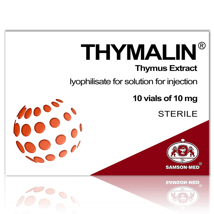
Thymalin® (Timalin) is a polypeptide medicine with a molecular weight of less than 10kDa containing an extract of young horned cattle thymus. Thymalin is a natural physiological immunostimulant. The medication is said to stabilize the immune response and regulate the T- and B-cell ratio.
Thymalin was isolated in 1974 and approved by the USSR Pharmacological Committee for clinical use in 1977. It is indicated for:
- Immunodeficiency states in adults and children (+6 months old);
- Acute and chronic viral and bacterial infections;
- Infectious purulent and septic processes;
- Suppression of immunity and hematopoiesis after chemotherapy or radiation therapy in patients with cancer;
- Persistent violations of the thymus function (radiation sickness, tumors of the thymus, surgical removal of the thymus).
According to the results of the latest clinical study of Dr Khavinson et al in 2020 Thymalin is recommended for use in the complex therapy of coronavirus infection and in vaccination against COVID-19. [21]
Thymogen
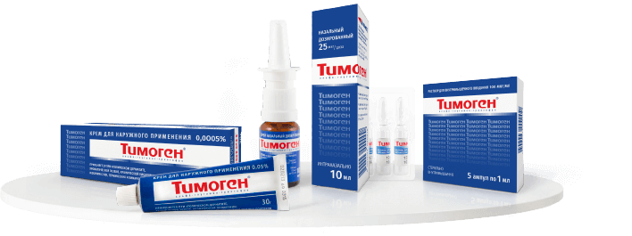
Thymogen® was originally isolated from Thymalin. It is a peptide immunomodulator containing chemically synthesized dipeptide Glu-Trp (glutamyl-tryptophan) equal to a natural compound of thymus extract. Thymogen is a versatile preparation that has several dosage forms available:
- Its spray form is usually used as a prophylactic and treatment of acute and chronic non-specific lung diseases.
- The injection form is used for the treatment of various acute and chronic viral and bacterial infections that are accompanied by weakened immunity.
- The cream form is used for the treatment of dermatitis and as a regeneration aid for the treatment of wounds and scars.
Thymogen was shown to be an effective immunomodulator as part of complex therapy of viral hepatitis, tuberculosis, pseudotuberculosis, influenza and acute respiratory viral infection (ARVI), papillomatosis and sepsis.
Thymogen improves patients’ general condition and ensures faster biochemical and immune restoration when used in the acute period of intestinal amebiasis, typhoid fever, acute dysentery, erysipelas, diphtheria, brucellosis, and hemorrhagic fever with renal syndrome. As a result of the use of the preparation, the incidence of prolonged recurrent and chronic disease is reduced.
Cytovir-3
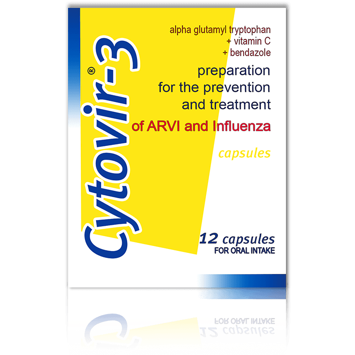
Cytovir®-3 is an immunomodulator, the purpose of which is to normalize the immune status. Cytovir-3 contains three active ingredients:
- Sodium thymogen (alpha glutamyl tryptophan), which helps to optimize the cellular and humoral links of immunity, as well as form protection when a low dose of the pathogen enters the body;
- Bendazol hydrochloride (dibazol) , which has the properties of a mild inducer of endogenous interferon and also modulates the functional activity of cells of the phagocytic-macrophage link;
- Ascorbic acid, which enhances the action of the other two components by activating the nonspecific defense system.
The basic principle of the complex is bioregulation, in which every component enhances the immunomodulatory potential of the other two. Together, they provide the immunostimulating effect.
Cytovir-3 is prescribed in the early stages of influenza and ARVI. According to the producer it:
- Strengthens the barrier function of the upper and lower respiratory airways;
- Specifically inhibits the multiplication of the SARS-CoV-2 virus, which is the causative agent of the new coronavirus infection COVID-19 (in vitro) [22] (Decree of the Ministry of Health of the Russian Federation No. 20-3-4144274/ID/IZM);
- Maintains the required level of the body’s own interferon, helping to fight viruses;
- Increases antioxidant protection of cells and tissues.
There are three forms of the drug: capsules for adults and children from 6 y.o., and powder and syrup for children from 1 y.o. because often infants find it difficult to swallow capsules.
References
- VG Morozov, VKh Khavinson (1973). Effect of substances isolated from the hypothalamus on immunogenesis and morphological composition of blood. 305 https://khavinson.info/ru/publications
- VG Morozov, VKh Khavinson (1974). Effect of epiphysis extract on the course of experimental tumors and leukemias. https://khavinson.info/ru/publications
- VG Morozov, VKh Khavinson (1978). Characteristics and study of the mechanism of action of thymus factor (thymarin). https://khavinson.info/ru/publications
- VG Morozov, VKh Khavinson (1981). Release of polypeptides from bone marrow, lymphocytes and thymus, which regulate the processes of intercellular cooperation in the immune system. https://khavinson.info/ru/publications
- VG Morozov, VKh Khavinson (1981). Isolation, purification, and identification of an immunomodulatory polypeptide contained in calf and human thymus. https://khavinson.info/ru/publications
- VG Morozov, VKh Khavinson (1983). New class of biological regulators of multicage systems – cytomedins. https://khavinson.info/assets/files/russ/1983-morozov_khavinson.pdf
- R. Gillemin (1982). Brain as an endocrine organ. https://arar.sci.am/dlibra/publication/290303/edition/266527/content
- VN Kalyunov (1984). Nerve tissue growth factor. https://opac.flib.sci.am/cgi-bin/koha/opac-detail.pl?biblionumber=69837
- VG Morozov, VKh Khavinson (1985). The role of cellular mediators (cytomedines) in the regulation of genetic activity. https://khavinson.info/assets/files/russ/1985-morozov_khavinson.pdf
- NP Prokopenko, RA Tigranian et al (1989). Characteristics of the acidic extract from the epiphysis and its fractions. https://pubmed.ncbi.nlm.nih.gov/2815684/
- GI Chipens, NI Veretennikova et al (1990). Structural basis of the action of peptide and protein immunoregulators. https://search.rsl.ru/ru/record/01001558284
- DD Wied (1990). Neuropeptides: Basics And Perspectives. https://www.goodreads.com/book/show/4194885-neuropeptides
- PK Klimov, GM Barashkova (1991). Physiology of the stomach. Mechanisms of regulation. https://www.ozon.ru/product/fiziologiya-zheludka-mehanizmy-regulyatsii-139626200/features/
- VN Anisimov, VKh Khavinson, VG Morozov (1993). The role of epiphysis peptides in homeostasis regulation: 20-year research experience. https://khavinson.info/ru/publications
- BI Kuznik, VKh Khavinson, VG Morozov (1995). Cytomedins and their role in the regulation of physiological functions. https://khavinson.info/ru/publications
- GN Akoev, NI Chalisova (1997). Neurotrophic regulation of nervous tissue. https://booksee.org/book/480818
- BI Kuznik, VKh Khavinson, VG Morozov (1998). CYTOMEDINS. 25 Years Of Experimental & Clinical Study. https://khavinson.info/ru/publications
- EG Elyashevich, ES Danchenko (2012). Alexander Vasilievich Poehl – great scientist. Pharmacist of the new time. https://www.elib.vsmu.by/bitstream/123/5592/1/vf_2012_2_50-55.pdf
- VS Saenko, DG Tzarichenko, SV Pesegov (2015). Cytomedins in the therapy of prostate diseases. https://lib.medvestnik.ru/articles/Citomediny-v-terapii-zabolevanii-prostaty.html
- OA Gomazkov (2011). Aging of the brain and neurotrophins. https://www.ibmc.msk.ru/content/monography/GomazkovOA3.pdf
- Khavinson V. Kh., Volchkov V.A. et al (2020). Effect of Thymalin on adaptive immunity in complex therapy for patients with Covid-19. https://khavinson.info/assets/files/russ/2020-khavinson_kuznik_volshkov.pdf
- Smirnov VS, Leneva IA et al (2021). Possibilities of Suppressing The Cytopathogenic Effect of SARS-CoV-2 Coronavirus According to The Results of the Antiviral Activity of Cytovir®-3 In Vitro Study. https://cytomed.ru/wp-content/uploads/2021/10/2vozmozhnosti-podavleniya-sars-cov-2.pdf


